


CONTACT
Dr. Nagaraj Balasubramanian
Dr. Homi Bhabha Road,
Pashan, Pune : 411008,
Maharashtra, INDIA
Tel No. : +91 (20) 25908202
Email. : adhesion.lab(at)gmail.com

RESEARCH INTEREST OF THE LAB
We are interested in understanding how the cell-matrix microenvironment affects what cells do. Cell-matrix interactions and their regulation of membrane trafficking, organelle architecture and signalling. Further, our studies test how this regulation is deregulated cancers to make cell anchorage-independent. We are aiming to target these deregulated pathways to restore anchorage-dependence in cancers. We also study how adhesion-dependent trafficking and signalling is different in 2D vs 3D microenvironment and how cells interact and behave with implants in a 3D microenvironment.


Congratulations
Dr. Kajal Singh
On successfully defending your thesis. 9th Graduating student from the lab.


Congratulations
Dr. Debasmita Mazumdar
On successfully defending your thesis. 10th Graduating student from the lab.

Congratulations
Dr. Arnav Saha
On successfully defending your thesis. 12th Graduating student from the lab.

Congratulations
Dr. Antara Chakraborty
On successfully defending your thesis. 11th Graduating student from the lab.
.png)


In MDAMB231 cells, the receptor tyrosine kinase AXL (magenta) localises to membrane ruffles (cyan overlayed image), and GM130-labelled Golgi (green), from the paper by Joshi et al. The overlay of the cyan image onto the membrane ruffles was performed using an image-editing tool.
(image Credit Arnav) - June 2026
NEW LAB PUBLICATIONS
click to open


LAB JOURNAL COVERS
click to open
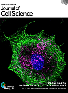

Oct 2024
Antara’s winning image ‘Breaking symmetry’ is the cover image for Journal of Cell Science’s Special Issue: Imaging Cell Architecture and Dynamics.
The image shows a mouse embryonic fibroblast labelled with phalloidin (actin – green), acetylated tubulin antibody (magenta) and DAPI (DNA – blue).
Image Credit : Antara


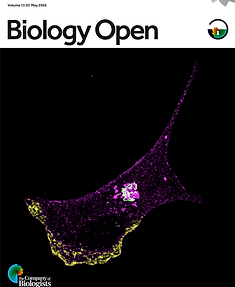
Jan 2026
The image shows a wild-type mouse embryonic fibroblast treated with ROCK inhibitor Y-27632 and embedded in 1.0 mg/mL 3D collagen gel. The cell is labeled with phalloidin for actin (purple), and the collagen is imaged by reflectance (yellow).
Image credit : Debasmita
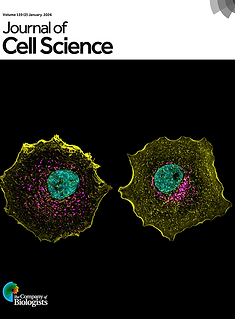

Jan 2026
Differential Golgi phenotypes in breast cancer cells. MCF7 cell populations exhibit contrasting Golgi organisations, with either a disorganised Golgi (as seen on the left) or a more compact organised Golgi (as seen on the right). Phalloidin-labelled actin (yellow), DAPI-stained nuclei (cyan) and GalTase–RFP-labelled trans-Golgi (magenta) reveal the differential Golgi organisation in MCF7 cells.
Image Credit : Arnav
PAST and CURRENT FUNDERS OF THE LAB


